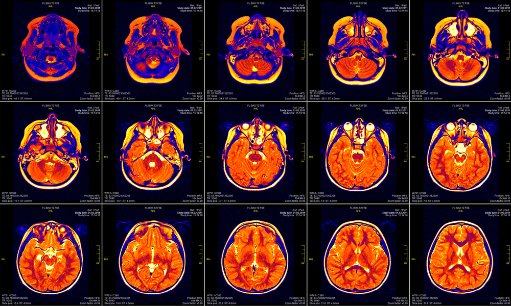
In medicine, imaging is forming a picture of a particular section the internal body structure, usually as a tool for diagnostic and treatment purposes. The materials used in this field commonly serve not only as imaging contrast agents but also as drug delivery systems.
Materials:
- Single-walled carbon nanohorns (SWCNHs)
These nanostructures were described as dahlia-like clusters of many horn-shaped sheaths of single-walled graphene sheets that were synthesized via CO2 laser ablation of carbon at room temperature with no metal catalyst.1 The cells from mice treated with SWCNHs were observed by a scanning probe microscopy technique called scanning near field ultrasonic holography, or simply, nanomechanical holography.2
- Ultra pH-sensitive (UPS) nanoprobes
These are polymeric, micelle-based nanoprobes that are non-toxic, fluorescent and highly responsive to pH so that in circulation they are silent (or stay “OFF” at normal pH 7.4) but are activated over 300-fold upon reaching the acidic extracellular tumor milieu (pH 6.5-6.8) and the endocytic environment (pH 5.0-6.0) of the cancer cells after receptor-mediated uptake. Basically, these series of UPS nanoprobes tested in this study consist of 3 components: a pH-sensitive core, a fluorophore (e.g., tetramethyl rhodamine, cyanine dyes, etc.), and a targeting unit (i.e., a short peptide sequence such as Arg-Gly-Asp).3
- Quantum dot bioconjugates (QDs)
Quantum dots have been shown to be ideal for deep tissue imaging due to their unique spectroscopic properties. This was demonstrated when near-infrared (NIR) CdTe(CdSe) core(shell) type II quantum dots with oligomeric phosphine coating for hydrophilicity were applied to sentinel lymph-node mapping in cancer surgeries in mice and pigs.4 Core-shell CdSe−ZnS quantum dots with various surface coating-dependent in vivo localization have also been utilized as noninvasive imaging contrast agents that were successfully detected via fluorescence imaging of living animals, by necropsy, by frozen tissue sections for optical microscopy, and by electron microscopy.5
- Carbon nanotubes (CNTs)
- Pristine CNTs – simplest form, e., no surface modification. The biodistribution single-walled carbon nanotubes (SWNTs) in mice was studied by using the skeleton 13C-enriched SWNTs and isotope ratio mass spectroscopy. Detection of SWNTs within the tissues were also analyzed by scanning TEM to explore a possible mechanism for uptake.6
- Surface coated CNTs – non-covalent surface modification. Such coatings has enhanced its dispersion and include lipid-polyethylene glycol (PEG) conjugates7, copolymers8, surfactants9, and DNA.10
- Functionalized CNTs – covalent surface modification.Nanomaterials used for different types of imaging technology for macrophages.11
- Magnetic resonance imaging (MRI) – biocompatible magnetic nanomaterials are usually made up of a magnetic core and a hydrophilic surface coating. Commonly used core materials include (1) monocrystalline iron oxide nanoparticles (MION) such as magnetite (Fe3O4) and maghemite (γ-Fe2O3), (2) cannonballs or elemental iron cores, (3) doped iron oxide cores, such as with Mn, and (4) pomegranate or multi-assembly cores in a silica coating. Solubilizing coatings typically involves hydrophilic polymers, g., dextrans, chitosan, starches, polyvinyl alcohol, polymethyl methacrylate, polyethylene glycol, polylactic-co-glycolic acid, polyvinylpyrrolidone, polyacrylic acid, etc.
- Positron emission tomography (PET) – nanoparticle-based PET agents contain materials such as modified dextrans and graft copolymers and are imaged with radiotracers or isotopes with different half-life depending on the circulation time: radioisotopes with long half-life (89Zr, 64Cu) for long circulating nanoparticles while radioisotopes with short half-life (18F) for rapidly excreted nanoparticles.
- Fluorescent nanomaterials – techniques such as microscopy, flow cytometry, endoscopy and intraoperative imaging require nanoparticles with affinity for macrophages which are generally divided into two categories: (1) fluorochrome-labeled polymeric nanoparticles, and (2) surface-stabilized quantum dots.
- X-ray computed tomography – this type of technology entails high (molar range) concentrations of absorbent nanoparticles and only very few were identified as useful, among which is the N1177 (iodinated emulsified suspension of a derivative of X-ray contrast agent diatrizoic acid) and PEGylated gold nanoparticles.
YouTube video: https://www.youtube.com/watch?v=SaqWPBMrOWI
SINAInnovations – Breakthroughs in Material Science, Nanotechnology and Imaging. Published on Nov. 20, 2014. Icahn School of Medicine at Mount Sinai (2nd of 3 video record). Guest Speaker: Angela M. Belcher (Chair, Dept of Biologica Engineering, MIT). Project: M13 phage was genetically engineered to bind SWNTs that fluoresce at NIR-II. This probe targeted SPARC-expressing ovarian tumor and was shown to be more fluorescently stable than the fluorescein derivative FITC. Enhanced tumor detection with mm to sub-mm resolution was made, and the study successfully demonstrated that the probe can be utilized both as a diagnostic method and as a surgical tool for ovarian cancer.
References:
- Iijima, S., Yudasaka, M., Yamada, R., Bandow, S., Suenaga, K., Kokai, F., Takahashi, K. (1999) Nano-aggregates of single-walled graphitic carbon nano-horns. Chemical Physics Letters 309:165-170.
- Tetard, L., Passian, A., Venmar, K. T., Lynch, R. M., Voy, B. H., Shekhawat, G., Dravid, V. P., Thundat, T. (2008) Imaging nanoparticles in cells by nanomechanical holography. Nature Nanotechnology 3:501-505.
- Wang, Y., Zhou, K., Huang, G., Hensley, C., Huang, X., Xinpeng, M., Zhao, T., Sumer, B. D., DeBerardinis R. J., Gao, J. (2014) A nanoparticle-based strategy for the imaging of a broad range of tumors by nonlinear amplification of microenvironment signals. Nature Materials 13:204-212.
- Kim, S., Lim, Y. T., Soltesz, E. G., De Grand, A. M., Lee, J., Nakayama, A., Parker, J. A., Mihaljevic T., Laurence R. G., Dor, D. M., Cohn, L. H., Bawendi, M. G., Frangioni J. V. (2004) Near-infrared fluorescent type II quantum dots for sentinel lymph node mapping. Nature Biotechnology 22:93-97.
- Ballou, B., Lagerhorn C., Ernst, L. A., Bruchez, M. P., Waggoner, A. S. (2004) Noninvasive imaging of quantum dots in mice. Bioconjugate Chemistry 15:79-86.
- Yang, S., Guo, W., Lin, Y., Deng, X., Wang, H., Sun, H., Liu, Y., Wang, X., Wang, W., Chen, M., Huang, Y., Sun, Y. (2007) Biodistribution of pristine single-walled carbon nanotubes in vivo. Journal of Physical Chemistry C 111:17761-17764.
- Liu, Z., Cai, W., He, L., Nakayama, N., Chen, K., Sun, X., Chen, X., Dai, H. (2007) In vivo biodistribution and highly efficient tumour targeting of carbon nanotubes in mice. Nature Nanotechnology 2:47-52.
- Ali-Boucetta, H., Al-Jamal, K. T., McCarthy, D., Prato, M., Bianco, A., Kostarelos, K. (2008) Multiwalled carbon nanotube-doxorubicin supramolecular complexes for cancer therapeutics. Chemical Communications 459-461.
- Gannon, C. J., Cherukuri, P., Yakobson, B. I., Cognet, L., Kanzius, J. S., Kitrell, C., Weisman, B., Pasquali, M., Schmidt, H. K., Smalley, R. E., Curley, S. A. (2007) Carbon nanotube-enhanced thermal destruction of cancer cells in a noninvasive radiofrequency field. Cancer 110:2654-2665.
- Zheng, M., Jagota, A., Semke, E. D., Diner, B. A., Mclean R. S., Lustig, S. R., Richardson, R. E., Tassi, N. G. (2003) DNA-assisted dispersion and separation of carbon nanotubes. Nature Materials 2:338-342.
- Weissleder, R., Nahrendorf, M., Pittet, M. J. (2014) Imaging macrophages with nanoparticles. Nature Materials 13:125-138.
